
Question: From NCERT NEET [Difficult level:Easy]
In human child, sex is determined by
1. Size and number of sperms in semen
2. Size of egg to be fertilized
3. Sex chromosomes of father
4. Sex chromosomes of mother
Subtopic: Sex Determination |
Answer ▽
3. Sex chromosomes of father
5.6.1 Sex Determination in Humans
It has already been mentioned that the sex determining mechanism in
case of humans is XY type. Out of 23 pairs of chromosomes present,
22 pairs are exactly same in both males and females; these are the
autosomes. A pair of X-chromosomes are present in the female, whereas
the presence of an X and Y chromosome are determinant of the male
characteristic. During spermatogenesis among males, two types of
gametes are produced. 50 per cent of the total sperm produced carry
the X-chromosome and the rest 50 per cent has Y-chromosome besides
the autosomes. Females, however, produce only one type of ovum with
an X-chromosome. There is an equal probability of fertilisation of the
ovum with the sperm carrying either X or Y chromosome. In case the
ovum fertilises with a sperm carrying X-chromosome the zygote develops
into a female (XX) and the fertilisation of ovum with Y-chromosome
carrying sperm results into a male offspring. Thus, it is evident that it
is the genetic makeup of the sperm that determines the sex of the child.
It is also evident that in each pregnancy there is always 50 per cent
probability of either a male or a female child. It is unfortunate that in
our society women are blamed for giving birth to female children and
have been ostracised and ill-treated because of this false notion
⬆️Prev____@organised notes_____Next⬇️
Question: From NCERT NEET [Difficult level:Easy]
Which of the following is a recessive trait for a character choosen by Mendel in garden pea?
(1) Violet flower color
(2) Yellow pod color
(3) Axial flower position
(4) Tall stem height
Subtopic: Introduction to Genetics:
Answer ▽
(2) Yellow pod color

⬆️Prev____@organised notes_____Next⬇️
Question: From NCERT NEET [Difficult level:Easy]
What is incorrect for Hemophilia?
1. In this disease, a single protein that is a part of the cascade of proteins involved in the clotting of blood is affected.
2. In an affected indlvidual a simple cut will result in non-stop bleeding.
3. The heterozygous female (carrier) for haemophilia may transmit the disease to sons.
4. The possibility of a female becoming a haemophilic is extremely rare because mother of such a female has to be hemophilic and the father should be a carrier.
Subtopic: Mendelian Disorders: Hemophilia |
Answer ▽
4. The possibility of a female becoming a haemophilic is extremely rare because mother of such a female has to be hemophilic and the father should be a carrier.
Haemophilia : This sex linked recessive disease, which shows its
transmission from unaffected carrier female to some of the male progeny
has been widely studied. In this disease, a single protein that is a part of
the cascade of proteins involved in the clotting of blood is affected. Due to
this, in an affected individual a simple cut will result in non-stop bleeding.
The heterozygous female (carrier) for haemophilia may transmit the disease
to sons. The possibility of a female becoming a haemophilic is extremely
rare because mother of such a female has to be at least carrier and the
father should be haemophilic (unviable in the later stage of life). The family
pedigree of Queen Victoria shows a number of haemophilic descendents
as she was a carrier of the disease.
⬆️Prev____@organised notes_____Next⬇️
Question: From NCERT NEET [Difficult level:Easy]
Sickle cell anaemia results from.
1. A chromosomal aberration
2. Non disjunction of autosome
3. A point mutation
4. Blood transfusion reaction
Answer ▽
3. A point mutation
Sickle-cell anaemia : This is an autosome linked recessive trait that can
be transmitted from parents to the offspring when both the partners are
carrier for the gene (or heterozygous). The disease is controlled by a single
pair of allele, HbA and HbS. Out of the three possible genotypes only
homozygous individuals for HbS (HbSHbS) show the diseased phenotype.
Heterozygous (HbAHbS) individuals appear apparently unaffected but they
are carrier of the disease as there is 50 per cent probability of transmission
of the mutant gene to the progeny, thus exhibiting sickle-cell trait
. The defect is caused by the substitution of Glutamic acid(Glu) by Valine (Val) at the sixth position of the beta globin chain of the
haemoglobin molecule. The substitution of amino acid in the globin
protein results due to the single base substitution at the sixth codon of
the beta globin gene from GAG to GUG. The mutant haemoglobin molecule
undergoes polymerisation under low oxygen tension causing the change
in the shape of the RBC.
⬆️Prev____@organised notes_____Next⬇️
Question: From NCERT NEET [Difficult level:Easy]
What is the mode of inheritance of phenylketonuria?
1. Autosomal recessive
2. Autosomal dominant
3. Sex linked recessive
4. Sex linked dominant
Answer ▽
1. Autosomal recessive
Phenylketonuria : This inborn error of metabolism is also inherited as
the autosomal recessive trait. The affected individual lacks an enzyme
that converts the amino acid phenylalanine into tyrosine. As a result of
this phenylalanine is accumulated and converted into phenylpyruvic acid
and other derivatives. Accumulation of these in brain results in mental
retardation. These are also excreted through urine because of its poor
absorption by kidney
⬆️Prev____@organised notes_____Next⬇️
Question: From NCERT NEET [Difficult level:Easy]
Failure of cytokinesis after telophase stage of cell divislon results in an increase in a whole set of chromosomes in an organism and, this phenomenon is known as:
1. Aneuploidy
2. Translocation
3. Polyploidy.
4. Inversion
Answer ▽
3. Polyploidy.
Chromosomal Disorders
The chromosomal disorders on the other hand are caused due to absence
or excess or abnormal arrangement of one or more chromosomes.
Failure of segregation of chromatids during cell division cycle results
in the gain or loss of a chromosome(s), called aneuploidy. For example,
Down’s syndrome results in the gain of extra copy of chromosome 21.
Similarly, Turner’s syndrome results due to loss of an X chromosome in
human females. Failure of cytokinesis after telophase stage of cell division
results in an increase in a whole set of chromosomes in an organism and,
this phenomenon is known as polyploidy. This condition is often seen in
plants.
Down’s Syndrome :
The cause of this genetic disorder
is the presence of an additional copy of the
chromosome number 21 (trisomy of 21). This disorder
was first described by Langdon Down (1866). The
affected individual is short statured with small round
head, furrowed tongue and partially open mouth
(Figure 5.16). Palm is broad with characteristic palm
crease. Physical, psychomotor and mental
development is retarded.
Klinefelter’s Syndrome :
This genetic disorder is also
caused due to the presence of an additional copy of Xchromosome
resulting into a karyotype of 47, XXY.
Such an individual has overall masculine development,
however, the feminine development (development
of breast, i.e., Gynaecomastia) is also expressed
(Figure 5.17 a). Such individuals are sterile.
Turner’s Syndrome :
Such a disorder is caused due
to the absence of one of the X chromosomes, i.e., 45 with X0, Such females
are sterile as ovaries are rudimentary besides other features including
lack of other secondary sexual characters .
⬆️Prev____@organised notes_____Next⬇️
Question: From NCERT NEET [Difficult level:Easy]
Which of the following is not a feature of Down's Syndrome?
1. It is caused by a non-disjunction in an autosome
2. The affected individual has trisomy of chromosome 21
3. The aflected individual has a characteristic simian palmar crease
4. The mental development of affected individual is normal
Answer ▽
1. It is caused by a non-disjunction in an autosome
Down’s Syndrome :
The cause of this genetic disorder
is the presence of an additional copy of the
chromosome number 21 (trisomy of 21). This disorder
was first described by Langdon Down (1866). The
affected individual is short statured with small round
head, furrowed tongue and partially open mouth
(Figure 5.16). Palm is broad with characteristic palm
crease. Physical, psychomotor and mental
development is retarded.
Klinefelter’s Syndrome :
This genetic disorder is also
caused due to the presence of an additional copy of Xchromosome
resulting into a karyotype of 47, XXY.
Such an individual has overall masculine development,
however, the feminine development (development
of breast, i.e., Gynaecomastia) is also expressed
(Figure 5.17 a). Such individuals are sterile.
Turner’s Syndrome :
Such a disorder is caused due
to the absence of one of the X chromosomes, i.e., 45 with X0, Such females
are sterile as ovaries are rudimentary besides other features including
lack of other secondary sexual characters .
⬆️Prev____@organised notes_____Next⬇️
Question: From NCERT NEET [Difficult level:Easy]
The two alleles of a gene pair are located on:
1. Homologous sites on homologous chromosomes
2. Heterologous sites on homologous chromosomes
3. Homologous sites on heterologous chromosomes
4. Heterologous sites on heretologous chromosomes
Answer ▽
1. Homologous sites on homologous chromosomes
In 1900, three Scientists (de Vries, Correns and von Tschermak)
independently rediscovered Mendel’s results on the inheritance of
characters. Also, by this time due to advancements in microscopy that
were taking place, scientists were able to carefully observe cell division.
This led to the discovery of structures in the nucleus that appeared to
double and divide just before each cell division. These were called
chromosomes (colored bodies, as they were visualised by staining). By
1902, the chromosome movement during meiosis had been worked out.
Walter Sutton and Theodore Boveri noted that the behaviour of
chromosomes was parallel to the behaviour of genes and used
chromosome movement (Figure 5.8) to explain Mendel’s laws (Table 5.3).
Recall that you have studied the behaviour of chromosomes during mitosis
(equational division) and during meiosis (reduction division). The
important things to remember are that chromosomes as well as genes
occur in pairs. The two alleles of a gene pair are located on homologous
sites on homologous chromosomes.
Additional-
During Anaphase of meiosis I, the two chromosome pairs can align at
the metaphase plate independently of each other.
During Zygotene of meiosis 1 stage chromosomes start pairing together
and this process of association is called synapsis. Such paired
chromosomes are called homologous chromosomes.
Recombination between homologous
chromosomes is completed by the end of pachytene, leaving the
chromosomes linked at the sites of crossing over.
⬆️Prev____@organised notes_____Next⬇️
Question: From NCERT NEET [Difficult level:Easy]
Male heterogamety is not seen in:
1. Humans
2. Melandrium album
3. Birds
4. Fruit fly
Answer ▽
3. Birds
SEX DETERMINATION
The mechanism of sex determination has always been a puzzle before the
geneticists. The initial clue about the genetic/chromosomal mechanism
of sex determination can be traced back to some of the experiments carried
out in insects. In fact, the cytological observations made in a number of
insects led to the development of the concept of genetic/chromosomal
basis of sex-determination. Henking (1891) could trace a specific nuclear
structure all through spermatogenesis in a few insects, and it was also
observed by him that 50 per cent of the sperm received this structure
after spermatogenesis, whereas the other 50 per cent sperm did not receive
it. Henking gave a name to this structure as the X body but he could not
explain its significance. Further investigations by other scientists led to
the conclusion that the ‘X body’ of Henking was in fact a chromosome and that is why it was given the name
X-chromosome. It was also observed that in
a large number of insects the mechanism of
sex determination is of the XO type, i.e., all
eggs bear an additional X-chromosome
besides the other chromosomes
(autosomes). On the other hand, some of the
sperms bear the X-chromosome whereas
some do not. Eggs fertilised by sperm having
an X-chromosome become females and,
those fertilised by sperms that do not have
an X-chromosome become males. Do you
think the number of chromosomes in the
male and female are equal? Due to the
involvement of the X-chromosome in the
determination of sex, it was designated to
be the sex chromosome, and the rest of the
chromosomes were named as
autosomes.Grasshopper is an example of
XO type of sex determination in which the
males have only one X-chromosome besides
the autosomes, whereas females have a pair
of X-chromosomes.
These observations led to the
investigation of a number of species to
understand the mechanism of sex
determination.
In a number of other insects
and mammals including man, XY type of sex
determination is seen where both male and
female have same number of chromosomes.
Among the males an X-chromosome is
present but its counter part is distinctly
smaller and called the Y-chromosome.
Females, however, have a pair of Xchromosomes.
Both males and females bear
same number of autosomes. Hence, the males have autosomes plus XY,
while female have autosomes plus XX. In human beings and in
Drosophila the males have one X and one Y chromosome, whereas females
have a pair of X-chromosomes besides autosomes (Figure 5.12 a, b).
In the above description you have studied about two types of sex
determining mechanisms, i.e., XO type and XY type. But in both cases
males produce two different types of gametes, (a) either with or without
X-chromosome or (b) some gametes with X-chromosome and some with
Y-chromosome. Such types of sex determination mechanism is designated
to be the example of male heterogamety. In some other organisms, e.g.,
birds, a different mechanism of sex determination is observed (Figure
5.12 c). In this case the total number of chromosome is same in both
males and females. But two different types of gametes in terms of the sex chromosomes, are produced by females, i.e., female heterogamety. In
order to have a distinction with the mechanism of sex determination
described earlier, the two different sex chromosomes of a female bird has
been designated to be the Z and W chromosomes. In these organisms the
females have one Z and one W chromosome, whereas males have a pair of
Z-chromosomes besides the autosomes.
⬆️Prev____@organised notes_____Next⬇️
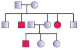
Question: From NCERT NEET [Difficult level:Easy]
The trait shown in the given pedigree chart is most likely a/an:
1. Autosomal recessive trait
2. Autosomal dominant trait
3. Sex linked recessive trait
4. Sex linked dominant trait
Answer ▽
1. Autosomal recessive trait

Pedigree Analysis
prevailing in the human society since long. This was
based on the heritability of certain characteristic
features in families. After the rediscovery of Mendel’s
work the practice of analysing inheritance pattern of
traits in human beings began. Since it is evident that
control crosses that can be performed in pea plant or
some other organisms, are not possible in case of
human beings, study of the family history about
inheritance of a particular trait provides an
alternative. Such an analysis of traits in a several of generations of a family
is called the pedigree analysis. In the pedigree analysis the inheritance
of a particular trait is represented in the family tree over generations.
In human genetics, pedigree study provides a strong tool, which is
utilised to trace the inheritance of a specific trait, abnormality or disease.
Some of the important standard symbols used in the pedigree analysis
have been shown in Figure 5.13.
As you have studied in this chapter, each and every feature in any
organism is controlled by one or the other gene located on the
DNA present in the chromosome. DNA is the carrier of genetic information. It is hence
transmitted from one generation to the other without any change or
alteration. However, changes or alteration do take place occasionally. Such
an alteration or change in the genetic material is referred to as mutation.
A number of disorders in human beings have been found to be associated
with the inheritance of changed or altered genes or chromosomes.
⬆️Prev____@organised notes_____Next⬇️
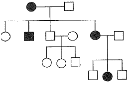
Question: From NCERT NEET [Difficult level:Easy]
The disease inheritance pattern exemplified in the given pedigree analysis can be :
1. Hemophilia
2. Red green colour blindness
3. Phenyl ketonuria
4. Polydactyly
Answer ▽
3. Phenyl ketonuria is correct.
Colour Blidness :
either red or green cone of eye resulting in failure to discriminate between
red and green colour. This defect is due to mutation in certain genes
present in the X chromosome. It occurs in about 8 per cent of males and
only about 0.4 per cent of females. This is because the genes that lead to
red-green colour blindness are on the X chromosome. Males have only
one X chromosome and females have two. The son of a woman who carries the gene has a 50 per cent chance of being colour blind. The mother is
not herself colour blind because the gene is recessive. That means that its
effect is suppressed by her matching dominant normal gene. A daughter
will not normally be colour blind, unless her mother is a carrier and her
father is colour blind.
Haemophilia :
transmission from unaffected carrier female to some of the male progeny
has been widely studied. In this disease, a single protein that is a part of
the cascade of proteins involved in the clotting of blood is affected. Due to
this, in an affected individual a simple cut will result in non-stop bleeding.
The heterozygous female (carrier) for haemophilia may transmit the disease
to sons. The possibility of a female becoming a haemophilic is extremely
rare because mother of such a female has to be at least carrier and the
father should be haemophilic (unviable in the later stage of life). The family
pedigree of Queen Victoria shows a number of haemophilic descendents
as she was a carrier of the disease.
Sickle-cell anaemia :
be transmitted from parents to the offspring when both the partners are
carrier for the gene (or heterozygous). The disease is controlled by a single
pair of allele, HbA and HbS. Out of the three possible genotypes only
homozygous individuals for HbS (HbSHbS) show the diseased phenotype.
Heterozygous (HbAHbS) individuals appear apparently unaffected but they
are carrier of the disease as there is 50 per cent probability of transmission
of the mutant gene to the progeny, thus exhibiting sickle-cell trait
(Figure 5.15). The defect is caused by the substitution of Glutamic acid (Glu) by Valine (Val) at the sixth position of the beta globin chain of the
haemoglobin molecule. The substitution of amino acid in the globin
protein results due to the single base substitution at the sixth codon of
the beta globin gene from GAG to GUG. The mutant haemoglobin molecule
undergoes polymerisation under low oxygen tension causing the change
in the shape of the RBC from biconcave disc to elongated sickle like
structure (Figure 5.15).
Phenylketonuria :
the autosomal recessive trait. The affected individual lacks an enzyme
that converts the amino acid phenylalanine into tyrosine. As a result of
this phenylalanine is accumulated and converted into phenylpyruvic acid
and other derivatives. Accumulation of these in brain results in mental
retardation. These are also excreted through urine because of its poor
absorption by kidney.
Thalassemia :
transmitted from parents to the offspring when both the partners are
unaffected carrier for the gene (or heterozygous). The defect could be due
to either mutation or deletion which ultimately results in reduced rate of
synthesis of one of the globin chains (a and b chains) that make up
haemoglobin. This causes the formation of abnormal haemoglobin
molecules resulting into anaemia which is characteristic of the disease.
Thalassemia can be classified according to which chain of the haemoglobin
molecule is affected. In a Thalassemia, production of a globin chain is
affected while in b Thalassemia, production of b globin chain is affected.
a Thalassemia is controlled by two closely linked genes HBA1 and HBA2
on chromosome 16 of each parent and it is observed due to mutation or
deletion of one or more of the four genes. The more genes affected, the
less alpha globin molecules produced. While b Thalassemia is controlled
by a single gene HBB on chromosome 11 of each parent and occurs due
to mutation of one or both the genes. Thalassemia differs from sickle-cell
anaemia in that the former is a quantitative problem of synthesising too
few globin molecules while the latter is a qualitative problem of
synthesising an incorrectly functioning globin.
Pedigree Analysis
prevailing in the human society since long. This was
based on the heritability of certain characteristic
features in families. After the rediscovery of Mendel’s
work the practice of analysing inheritance pattern of
traits in human beings began. Since it is evident that
control crosses that can be performed in pea plant or
some other organisms, are not possible in case of
human beings, study of the family history about
inheritance of a particular trait provides an
alternative. Such an analysis of traits in a several of generations of a family
is called the pedigree analysis. In the pedigree analysis the inheritance
of a particular trait is represented in the family tree over generations.
In human genetics, pedigree study provides a strong tool, which is
utilised to trace the inheritance of a specific trait, abnormality or disease.
Some of the important standard symbols used in the pedigree analysis
have been shown in Figure 5.13.
As you have studied in this chapter, each and every feature in any
organism is controlled by one or the other gene located on the
DNA present in the chromosome. DNA is the carrier of genetic information. It is hence
transmitted from one generation to the other without any change or
alteration. However, changes or alteration do take place occasionally. Such
an alteration or change in the genetic material is referred to as mutation.
A number of disorders in human beings have been found to be associated
with the inheritance of changed or altered genes or chromosomes.
⬆️Prev____@organised notes_____Next⬇️
Question: From NCERT NEET [Difficult level:Easy] NEET - 2015
A gene showing codominance has
(1) one allele dominant on the other
(2) alleles tightly linked on the same chromosome
(3) alleles that are recessive to each other
(4) both alleles independently expressed in the heterozygote
Answer ▽
(4) both alleles independently expressed in the heterozygote
Co-dominance
Till now we were discussing crosses where the F1 resembled either of the
two parents (dominance) or was in-between (incomplete dominance). But,
in the case of co-dominance the F1 generation resembles both parents. A
good example is different types of red blood cells that determine ABO
blood grouping in human beings. ABO blood groups are controlled by
the gene I. The plasma membrane of the red blood cells has sugar polymers
that protrude from its surface and the kind of sugar is controlled by the
gene. The gene (I) has three alleles IA, IB and i. The alleles IA and IB produce
a slightly different form of the sugar while allele i does not produce any
sugar. Because humans are diploid organisms, each person possesses
any two of the three I gene alleles. IA and IB are completely dominant over
i, in other words when IA and i are present only IA expresses (because i
does not produce any sugar), and when IB and i are present IB expresses.
But when IA and IB are present together they both express their own types
of sugars: this is because of co-dominance. Hence red blood cells have
both A and B types of sugars. Since there are three different alleles, there
are six different combinations of these three alleles that are possible, and
therefore, a total of six different genotypes of the human ABO blood types
(Table 5.2). How many phenotypes are possible?
⬆️Prev____@organised notes_____Next⬇️
Question: From NCERT NEET [Difficult level:Easy] NEET - 2017
Which one from those given below is the period of Mendel's hybridisation experiments?
(1) 1856 - 1863
(2) 1840 - 1850
(3) 1857 - 1869
(4) 1870 - 1877
Answer ▽
(1) 1856 - 1863
MENDEL’S LAWS OF INHERITANCE
It was during the mid-nineteenth century that
headway was made in the understanding of
inheritance. Gregor Mendel, conducted
hybridisation experiments on garden peas for
seven years (1856-1863) and proposed the
laws of inheritance in living organisms. During
Mendel’s investigations into inheritance
patterns it was for the first time that statistical
analysis and mathematical logic were applied
to problems in biology. His experiments had a
large sampling size, which gave greater
credibility to the data that he collected. Also,
the confirmation of his inferences from
experiments on successive generations of his
test plants, proved that his results pointed to
general rules of inheritance rather than being
unsubstantiated ideas. Mendel investigated
characters in the garden pea plant that were
manifested as two opposing traits, e.g., tall or
dwarf plants, yellow or green seeds. This
allowed him to set up a basic framework of
rules governing inheritance, which was
expanded on by later scientists to account for
all the diverse natural observations and the
complexity inherent in them.
Mendel conducted such artificial
pollination/cross pollination experiments
using several true-breeding pea lines. A true breeding
line is one that, having undergone
continuous self-pollination, shows the stable trait inheritance and
expression for several generations. Mendel selected 14 true-breeding pea
plant varieties, as pairs which were similar except for one character with
contrasting traits. Some of the contrasting traits selected were smooth or
wrinkled seeds, yellow or green seeds, inflated (full) or constricted green
or yellow pods and tall or dwarf plants.
⬆️Prev____@organised notes_____Next⬇️
Question: From NCERT NEET [Difficult level:Easy]
A tall true breeding garden pea plant is crossed with a dwarf true breeding garden pea plant. When the F1 plants were selfed the resulting genotypes were in the ratio of
(1) 1 : 2 : 1 :: Tall heterozygous : tall homozygous : Dwarf
(2) 3 : 1 :: Tall : Dwarf
(3) 3 : 1 :: Dwarf : Tall
(4) 1 : 2 : 1 :: Tall homozygous : Tall heterogygous : Dwarf
Answer ▽
(4) 1 : 2 : 1 :: Tall homozygous : Tall heterogygous : Dwarf
Though the F1
have a genotype of Tt, but the phenotypic character seen is ‘tall’. At F2,
3/4th of the plants are tall, where some of them are TT while others are
Tt. Externally it is not possible to distinguish between the plants with
the genotypes TT and Tt. Hence, within the genopytic pair Tt only one
character ‘T’ tall is expressed. Hence the character T or ‘tall’ is said to
dominate over the other allele t or ‘dwarf’ character. It is thus due to this
dominance of one character over the other that all the F1 are tall (though
the genotype is Tt) and in the F2 3/4th of the plants are tall (though
genotypically 1/2 are Tt and only 1/4th are TT). This leads to a phenotypic
ratio of 3/4th tall : (1/4 TT + 1/2 Tt) and 1/4th tt, i.e., a 3:1 ratio, but a
genotypic ratio of 1:2:1.
⬆️Prev____@organised notes_____Next⬇️
Question: From NCERT NEET [Difficult level:Easy] NEET - 2016
Pick out the correct statements.
I. Haemophilia is a sex-linked recessive disease
II. Down's syndrome is due to aneuploidy.
III. Phenylketonuria is an autosomal recessive gene disorder
IV. Sickle cell anaemia is an x - linked recessive gene disorder
(1) II and IV are correct
(2) I, III and IV are correct
(3) I, II and III are correct
(4) I and IV are correct
Answer ▽
(3) I, II and III are correct
Colour Blidness :
either red or green cone of eye resulting in failure to discriminate between
red and green colour. This defect is due to mutation in certain genes
present in the X chromosome. It occurs in about 8 per cent of males and
only about 0.4 per cent of females. This is because the genes that lead to
red-green colour blindness are on the X chromosome. Males have only
one X chromosome and females have two. The son of a woman
not herself colour blind because the gene is recessive. That means that its
effect is suppressed by her matching dominant normal gene. A daughter
will not normally be colour blind, unless her mother is a carrier and her
father is colour blind.
Haemophilia :
transmission from unaffected carrier female to some of the male progeny
has been widely studied. In this disease, a single protein that is a part of
the cascade of proteins involved in the clotting of blood is affected. Due to
this, in an affected individual a simple cut will result in non-stop bleeding.
The heterozygous female (carrier) for haemophilia may transmit the disease
to sons. The possibility of a female becoming a haemophilic is extremely
rare because mother of such a female has to be at least carrier and the
father should be haemophilic (unviable in the later stage of life). The family
pedigree of Queen Victoria shows a number of haemophilic descendents
as she was a carrier of the disease.
Sickle-cell anaemia :
be transmitted from parents to the offspring when both the partners are
carrier for the gene (or heterozygous). The disease is controlled by a single
pair of allele, HbA and HbS. Out of the three possible genotypes only
homozygous individuals for HbS (HbSHbS) show the diseased phenotype.
Heterozygous (HbAHbS) individuals appear apparently unaffected but they
are carrier of the disease as there is 50 per cent probability of transmission
of the mutant gene to the progeny, thus exhibiting sickle-cell trait
(Figure 5.15). The defect is caused by the substitution of Glutamic acid (Glu) by Valine (Val) at the sixth position of the beta globin chain of the
haemoglobin molecule. The substitution of amino acid in the globin
protein results due to the single base substitution at the sixth codon of
the beta globin gene from GAG to GUG. The mutant haemoglobin molecule
undergoes polymerisation under low oxygen tension causing the change
in the shape of the RBC from biconcave disc to elongated sickle like
structure (Figure 5.15).
Phenylketonuria :
the autosomal recessive trait. The affected individual lacks an enzyme
that converts the amino acid phenylalanine into tyrosine. As a result of
this phenylalanine is accumulated and converted into phenylpyruvic acid
and other derivatives. Accumulation of these in brain results in mental
retardation. These are also excreted through urine because of its poor
absorption by kidney.
Thalassemia :
transmitted from parents to the offspring when both the partners are
unaffected carrier for the gene (or heterozygous). The defect could be due
to either mutation or deletion which ultimately results in reduced rate of
synthesis of one of the globin chains (a and b chains) that make up
haemoglobin. This causes the formation of abnormal haemoglobin
molecules resulting into anaemia which is characteristic of the disease.
Thalassemia can be classified according to which chain of the haemoglobin
molecule is affected. In a Thalassemia, production of a globin chain is
affected while in b Thalassemia, production of b globin chain is affected.
a Thalassemia is controlled by two closely linked genes HBA1 and HBA2
on chromosome 16 of each parent and it is observed due to mutation or
deletion of one or more of the four genes. The more genes affected, the
less alpha globin molecules produced. While b Thalassemia is controlled
by a single gene HBB on chromosome 11 of each parent and occurs due
to mutation of one or both the genes. Thalassemia differs from sickle-cell
anaemia in that the former is a quantitative problem of synthesising too
few globin molecules while the latter is a qualitative problem of
synthesising an incorrectly functioning globin.
⬆️Prev____@organised notes_____Next⬇️
Question: From NCERT NEET [Difficult level:Easy] NEET - 2016
In a test cross involving F1 dihybrid flies, more parental-type offspring were produced than the recombinant type offspring. This indicates
(1) chromosomes failed to separate during meiosis
(2) the two genes are linked and present on the same chromosome
(3) both of the characters are controlled by more than one gene
(4) the two genes are located on two different chromosomes
Answer ▽
(2) the two genes are linked and present on the same chromosome
Linkage and Recombination
Morgan carried out several dihybrid crosses in Drosophila to study genes
that were sex-linked. The crosses were similar to the dihybrid crosses carried
out by Mendel in peas. For example Morgan hybridised yellow-bodied,
white-eyed females to brown-bodied, red-eyed males and intercrossed their
F1 progeny. He observed that the two genes did not segregate independently
of each other and the F2 ratio deviated very significantly from the 9:3:3:1
ratio (expected when the two genes are independent).
Morgan and his group knew that the genes were located on the X
chromosome (Section 5.4) and saw quickly that
when the two genes in a
dihybrid cross were situated on the same chromosome, the proportion
of parental gene combinations were much higher than the non-parental
type.
of the two genes and coined the term linkage to describe this physical
association of genes on a chromosome and the term recombination to
describe the generation of non-parental gene combinations (Figure 5.11).
Morgan and his group also found that even when genes were grouped
on the same chromosome, some genes were very tightly linked (showed
very low recombination) (Figure 5.11, Cross A) while others were loosely
linked (showed higher recombination) (Figure 5.11, Cross B). For
example he found that the genes white and yellow were very tightly linked
and showed only 1.3 per cent recombination while white and miniature
wing showed 37.2 per cent recombination. His student Alfred
Sturtevant used the frequency of recombination between gene pairs
on the same chromosome as a measure of the distance between genes
and ‘mapped’ their position on the chromosome. Today genetic maps
are extensively used as a starting point in the sequencing of whole
genomes as was done in the case of the Human Genome Sequencing
Project, described later.
⬆️Prev____@organised notes_____Next⬇️
Question: From NCERT NEET [Difficult level:Easy] NEET - 2016
Which of the following most appropriately describes haemophilia?
(1) X-linked recessive gene disorder
(2) Choromosomal disorder
(3) dominant gene disorder
(4) Recessive gene disorder
Answer ▽
(1) X-linked recessive gene disorder
Colour Blidness :
either red or green cone of eye resulting in failure to discriminate between
red and green colour. This defect is due to mutation in certain genes
present in the X chromosome. It occurs in about 8 per cent of males and
only about 0.4 per cent of females. This is because the genes that lead to
red-green colour blindness are on the X chromosome. Males have only
one X chromosome and females have two. The son of a woman
not herself colour blind because the gene is recessive. That means that its
effect is suppressed by her matching dominant normal gene. A daughter
will not normally be colour blind, unless her mother is a carrier and her
father is colour blind.
Haemophilia :
transmission from unaffected carrier female to some of the male progeny
has been widely studied. In this disease, a single protein that is a part of
the cascade of proteins involved in the clotting of blood is affected. Due to
this, in an affected individual a simple cut will result in non-stop bleeding.
The heterozygous female (carrier) for haemophilia may transmit the disease
to sons. The possibility of a female becoming a haemophilic is extremely
rare because mother of such a female has to be at least carrier and the
father should be haemophilic (unviable in the later stage of life). The family
pedigree of Queen Victoria shows a number of haemophilic descendents
as she was a carrier of the disease.
Sickle-cell anaemia :
be transmitted from parents to the offspring when both the partners are
carrier for the gene (or heterozygous). The disease is controlled by a single
pair of allele, HbA and HbS. Out of the three possible genotypes only
homozygous individuals for HbS (HbSHbS) show the diseased phenotype.
Heterozygous (HbAHbS) individuals appear apparently unaffected but they
are carrier of the disease as there is 50 per cent probability of transmission
of the mutant gene to the progeny, thus exhibiting sickle-cell trait
(Figure 5.15). The defect is caused by the substitution of Glutamic acid (Glu) by Valine (Val) at the sixth position of the beta globin chain of the
haemoglobin molecule. The substitution of amino acid in the globin
protein results due to the single base substitution at the sixth codon of
the beta globin gene from GAG to GUG. The mutant haemoglobin molecule
undergoes polymerisation under low oxygen tension causing the change
in the shape of the RBC from biconcave disc to elongated sickle like
structure (Figure 5.15).
Phenylketonuria :
the autosomal recessive trait. The affected individual lacks an enzyme
that converts the amino acid phenylalanine into tyrosine. As a result of
this phenylalanine is accumulated and converted into phenylpyruvic acid
and other derivatives. Accumulation of these in brain results in mental
retardation. These are also excreted through urine because of its poor
absorption by kidney.
Thalassemia :
transmitted from parents to the offspring when both the partners are
unaffected carrier for the gene (or heterozygous). The defect could be due
to either mutation or deletion which ultimately results in reduced rate of
synthesis of one of the globin chains (a and b chains) that make up
haemoglobin. This causes the formation of abnormal haemoglobin
molecules resulting into anaemia which is characteristic of the disease.
Thalassemia can be classified according to which chain of the haemoglobin
molecule is affected. In a Thalassemia, production of a globin chain is
affected while in b Thalassemia, production of b globin chain is affected.
a Thalassemia is controlled by two closely linked genes HBA1 and HBA2
on chromosome 16 of each parent and it is observed due to mutation or
deletion of one or more of the four genes. The more genes affected, the
less alpha globin molecules produced. While b Thalassemia is controlled
by a single gene HBB on chromosome 11 of each parent and occurs due
to mutation of one or both the genes. Thalassemia differs from sickle-cell
anaemia in that the former is a quantitative problem of synthesising too
few globin molecules while the latter is a qualitative problem of
synthesising an incorrectly functioning globin.
⬆️Prev____@organised notes_____Next⬇️
Question: From NCERT NEET [Difficult level:Easy] NEET - 2015
The term "linkage" was coined by :-
1. TH Morgan
2. T Boveri
3. G Mendel
4. W Sulton
Answer ▽
1. TH Morgan
Linkage and Recombination
Morgan carried out several dihybrid crosses in Drosophila to study genes
that were sex-linked. The crosses were similar to the dihybrid crosses carried
out by Mendel in peas. For example Morgan hybridised yellow-bodied,
white-eyed females to brown-bodied, red-eyed males and intercrossed their
F1 progeny. He observed that the two genes did not segregate independently
of each other and the F2 ratio deviated very significantly from the 9:3:3:1
ratio (expected when the two genes are independent).
Morgan and his group knew that the genes were located on the X
chromosome (Section 5.4) and saw quickly that
when the two genes in a
dihybrid cross were situated on the same chromosome, the proportion
of parental gene combinations were much higher than the non-parental
type.
of the two genes and coined the term linkage to describe this physical
association of genes on a chromosome and the term recombination to
describe the generation of non-parental gene combinations (Figure 5.11).
Morgan and his group also found that even when genes were grouped
on the same chromosome, some genes were very tightly linked (showed
very low recombination) (Figure 5.11, Cross A) while others were loosely
linked (showed higher recombination) (Figure 5.11, Cross B). For
example he found that the genes white and yellow were very tightly linked
and showed only 1.3 per cent recombination while white and miniature
wing showed 37.2 per cent recombination. His student Alfred
Sturtevant used the frequency of recombination between gene pairs
on the same chromosome as a measure of the distance between genes
and ‘mapped’ their position on the chromosome. Today genetic maps
are extensively used as a starting point in the sequencing of whole
genomes as was done in the case of the Human Genome Sequencing
Project, described later.
⬆️Prev____@organised notes_____Next⬇️
Question: From NCERT NEET [Difficult level:Easy] NEET - 2015
A pleiotropic gene
a. is expressed only in primitive plants
b. is a genet envolved during Pliocene
c. controls a trait only in combination 'Nith another gene
d. control multiple traits in an individual
Answer ▽
d. control multiple traits in an individual
PLEIOTROPY
We have so far seen the effect of a gene on a single phenotype or trait.
There are however instances where a single gene can exhibit multiple
phenotypic expression. Such a gene is called a pleiotropic gene. The
underlying mechanism of pleiotropy in most cases is the effect of a gene
on metabolic pathways which contribute towards different phenotypes.
An example of this is the disease phenylketonuria, which occurs in
humans. The disease is caused by mutation in the gene that codes for the
enzyme phenyl alanine hydroxylase (single gene mutation). This manifests
itself through phenotypic expression characterised by mental
retardation and a reduction in hair and skin pigmentation.
Additional confusing terms-
The chromosomal disorders on the other hand are caused due to absence
or excess or abnormal arrangement of one or more chromosomes.
Failure of segregation of chromatids during cell division cycle results
in the gain or loss of a chromosome(s), called aneuploidy. For example,
Down’s syndrome results in the gain of extra copy of chromosome 21.
Similarly, Turner’s syndrome results due to loss of an X chromosome in
human females. Failure of cytokinesis after telophase stage of cell division
results in an increase in a whole set of chromosomes in an organism and,
this phenomenon is known as polyploidy. This condition is often seen in
plants.
⬆️Prev____@organised notes_____Next⬇️
Question: From NCERT NEET [Difficult level:Easy] NEET - 2015
Alleles are
1. different phenotype
2. true breeding homozygotes
3. different molecular forms of a gene
4. heterozygotes
Answer ▽
3. different molecular forms of a gene
Alleles are variations or forms of a same gene located at a specific chromosomal site. Alleles are generally present in pairs and control the same character or trait. Therefore, we can say alleles are an alternative form of a gene.
A gene is a specific nucleotide sequence that encodes for a particular protein. Alleles are variations of the same gene. Alleles arise by mutation of either a single base pair or several hundred base pairs. They are present in pairs – one allele inherited from the father and the other from the mother. Alleles influence the same phenotype or character. For example, consider a phenotype – eye colour. Colour of the eye can be black, brown, blue or even green. Therefore, blue, black, brown and green are the alleles of the gene that influence eye colour. Thus, we can say that alleles are the different molecular forms of a gene.
Heterozygotes are organisms that contain two different alleles of a gene. The organism will be heterozygous for that particular gene. Heterozygosity is seen in diploid organisms, where one allele is received from the father and the other from the mother. Out of the two alleles present, one allele generally dominates the other one and is expressed in the off-spring. It is known as the dominant allele and the other allele, the recessive allele. Dominant traits are denoted by capital letters, while recessive ones by small letters. Thus, a heterozygote individual can be denoted as Aa.
Phenotypes are visible traits. They are the physical characteristics of an individual like, hair colour, eye colour, skin colour, etc. Phenotypes are controlled by alleles; however, they are not alleles.
Homozygotes are organisms that contain the same allele on both the homologous chromosomes. Homozygotes can be denoted by AA or aa. AA individuals are called homozygous dominants, since they contain two dominant alleles. While aa individuals are homozygous recessives, because they contain two recessive alleles. True breeding Homozygotes are purebred organisms that are homozygous for certain phenotypes, that always pass down the same trait to off-springs for many generations.
Therefore, from the above discussion it can be concluded that option (3) is correct.
Alleles are the different forms of a gene that occupy the same position on homologous chromosomes. Alleles are present in pairs and influence the same character. Alleles can be dominant or recessive. Dominant alleles obscure the expression of recessive alleles. Hair colour, skin colour, eye colour, different blood groups are all examples of alleles present in the population.
⬆️Prev____@organised notes_____Next⬇️
Question: From NCERT NEET [Difficult level:Easy] NEET - 2014
A human female with turner's syndrome
(1) has 45 chromosomes with XO
(2) has one additional X-chromosome
(3) exhibits male characters
(4) is able to produce children with normal husband
Answer ▽
(1) has 45 chromosomes with XO
Down’s Syndrome :
The cause of this genetic disorder
is the presence of an additional copy of the
chromosome number 21 (trisomy of 21). This disorder
was first described by Langdon Down (1866). The
affected individual is short statured with small round
head, furrowed tongue and partially open mouth
(Figure 5.16). Palm is broad with characteristic palm
crease. Physical, psychomotor and mental
development is retarded.
Klinefelter’s Syndrome :
This genetic disorder is also
caused due to the presence of an additional copy of X chromosome
resulting into a karyotype of 47, XXY.
Such an individual has overall masculine development,
however, the feminine development (development
of breast, i.e., Gynaecomastia) is also expressed
(Figure 5.17 a). Such individuals are sterile.
Turner’s Syndrome :
Such a disorder is caused due
to the absence of one of the X chromosomes, i.e., 45 with X0, Such females
are sterile as ovaries are rudimentary besides other features including
lack of other secondary sexual characters
⬆️Prev____@organised notes_____Next⬇️
Question: From NCERT NEET [Difficult level:Easy]
Which one of the following conditions correctly describes the manner of determining the sex in the given example?
(1) XO type of sex chromosomes determine male sex in grasshopper
(2) XO condition in humans as found in Turner syndromee, determines female sex
(3) Homozygous sex chromosomes (XX) produce male in Drosophila
(4) Homozygous sex chromosomes (ZZ) determine female sex in birds
Answer ▽
(1) XO type of sex chromosomes determine male sex in grasshopper
SEX DETERMINATION
The mechanism of sex determination has always been a puzzle before the
geneticists. The initial clue about the genetic/chromosomal mechanism
of sex determination can be traced back to some of the experiments carried
out in insects. In fact, the cytological observations made in a number of
insects led to the development of the concept of genetic/chromosomal
basis of sex-determination. Henking (1891) could trace a specific nuclear
structure all through spermatogenesis in a few insects, and it was also
observed by him that 50 per cent of the sperm received this structure
after spermatogenesis, whereas the other 50 per cent sperm did not receive
it. Henking gave a name to this structure as the X body but he could not
explain its significance. Further investigations by other scientists led to
the conclusion that the ‘X body’ of Henking was in fact a chromosome and that is why it was given the name
X-chromosome. It was also observed that in
a large number of insects the mechanism of
sex determination is of the XO type, i.e., all
eggs bear an additional X-chromosome
besides the other chromosomes
(autosomes). On the other hand, some of the
sperms bear the X-chromosome whereas
some do not. Eggs fertilised by sperm having
an X-chromosome become females and,
those fertilised by sperms that do not have
an X-chromosome become males. Do you
think the number of chromosomes in the
male and female are equal? Due to the
involvement of the X-chromosome in the
determination of sex, it was designated to
be the sex chromosome, and the rest of the
chromosomes were named as
autosomes.Grasshopper is an example of
XO type of sex determination in which the
males have only one X-chromosome besides
the autosomes, whereas females have a pair
of X-chromosomes.
These observations led to the
investigation of a number of species to
understand the mechanism of sex
determination.
In a number of other insects
and mammals including man, XY type of sex
determination is seen where both male and
female have same number of chromosomes.
Among the males an X-chromosome is
present but its counter part is distinctly
smaller and called the Y-chromosome.
Females, however, have a pair of Xchromosomes.
Both males and females bear
same number of autosomes. Hence, the males have autosomes plus XY,
while female have autosomes plus XX. In human beings and in
Drosophila the males have one X and one Y chromosome, whereas females
have a pair of X-chromosomes besides autosomes (Figure 5.12 a, b).
In the above description you have studied about two types of sex
determining mechanisms, i.e., XO type and XY type. But in both cases
males produce two different types of gametes, (a) either with or without
X-chromosome or (b) some gametes with X-chromosome and some with
Y-chromosome. Such types of sex determination mechanism is designated
to be the example of male heterogamety. In some other organisms, e.g.,
birds, a different mechanism of sex determination is observed (Figure
5.12 c). In this case the total number of chromosome is same in both
males and females. But two different types of gametes in terms of the sex chromosomes, are produced by females, i.e., female heterogamety. In
order to have a distinction with the mechanism of sex determination
described earlier, the two different sex chromosomes of a female bird has
been designated to be the Z and W chromosomes. In these organisms the
females have one Z and one W chromosome, whereas males have a pair of
Z-chromosomes besides the autosomes.
⬆️Prev____@organised notes_____Next⬇️
Question: From NCERT NEET [Difficult level:Easy] NEET - 2010
Which one of the following cannot be explained on the basis of Mendel's Law of Dominance?
(1) The discrete unit controlling a particular character is called a factor
(2) Out of one pair of factors one is dominant, and the other is recessive
(3) Alleles do not show any blendings and both the characters recover as such in F2 generation.
(4) Factors occur in pairs
Answer ▽
(3) Alleles do not show any blendings and both the characters recover as such in F2 generation.
Law of Dominance
(i) Characters are controlled by discrete units called factors.
(ii) Factors occur in pairs.
(iii) In a dissimilar pair of factors one member of the pair dominates
(dominant) the other (recessive).
The law of dominance is used to explain the expression of only one of
the parental characters in a monohybrid cross in the F1 and the expression
of both in the F2. It also explains the proportion of 3:1 obtained at the F2.
Law of Segregation
This law is based on the fact that the alleles do not show any blending
and that both the characters are recovered as such in the F2 generation
though one of these is not seen at the F1 stage. Though the parents contain
two alleles during gamete formation, the factors or alleles of a pair segregate
from each other such that a gamete receives only one of the two factors.
Of course, a homozygous parent produces all gametes that are similar
while a heterozygous one produces two kinds of gametes each having
one allele with equal proportion.
Law of Independent Assortment
In the dihybrid cross (Figure 5.7), the phenotypes round, yellow;
wrinkled, yellow; round, green and wrinkled, green appeared in the
ratio 9:3:3:1. Such a ratio was observed for several pairs of characters
that Mendel studied.
The ratio of 9:3:3:1 can be derived as a combination series of 3 yellow:
1 green, with 3 round : 1 wrinkled. This derivation can be written
as follows:
(3 Round : 1 Wrinkled) (3 Yellow : 1 Green) = 9 Round, Yellow : 3
Wrinkled, Yellow: 3 Round, Green : 1 Wrinkled, Green
Based upon such observations on dihybrid crosses (crosses between
plants differing in two traits) Mendel proposed a second set of generalisations
that we call Mendel’s Law of Independent Assortment. The law states that
‘when two pairs of traits are combined in a hybrid, segregation of one pair
of characters is independent of the other pair of characters’.
⬆️Prev____@organised notes_____Next⬇️
Question: From NCERT NEET [Difficult level:Easy] NEET - 2010
Which one of the following symbols and its representation, used in human pedigree analysis is correct?
(1) 
(2) 
(3) 
(4) 
Answer ▽
(1)

⬆️Prev____@organised notes_____Next⬇️
Question: From NCERT NEET [Difficult level:Easy] NEET - 2008
Which one of the following condition in human is correctly matched with its chromosomal abnormality/linkage?
(1) Klinefelter's syndrome—44 autosomes + XXY
(2) Colourblindness —Y-linked
(3) Erythroblastosis foetalis— X-linked
(4) Down syndrome—44 autosomes + XO
Answer ▽
(1) Klinefelter's syndrome—44 autosomes + XXY
Down’s Syndrome :
The cause of this genetic disorder
is the presence of an additional copy of the
chromosome number 21 (trisomy of 21). This disorder
was first described by Langdon Down (1866). The
affected individual is short statured with small round
head, furrowed tongue and partially open mouth
(Figure 5.16). Palm is broad with characteristic palm
crease. Physical, psychomotor and mental
development is retarded.
Klinefelter’s Syndrome :
This genetic disorder is also
caused due to the presence of an additional copy of X chromosome
resulting into a karyotype of 47, XXY.
Such an individual has overall masculine development,
however, the feminine development (development
of breast, i.e., Gynaecomastia) is also expressed
(Figure 5.17 a). Such individuals are sterile.
Turner’s Syndrome :
Such a disorder is caused due
to the absence of one of the X chromosomes, i.e., 45 with X0, Such females
are sterile as ovaries are rudimentary besides other features including
lack of other secondary sexual characters
⬆️Prev____@organised notes_____Next⬇️
Question: From NCERT NEET [Difficult level:Easy] NEET - 2007
Inheritance of skin colour in humans is an example of :
(1) chromosomal aberration
(2) point mutation
(3) polygenic inheritance
(4) codominance
Answer ▽
(3) polygenic inheritance
POLYGENIC INHERITANCE
Mendel’s studies mainly described those traits that have distinct alternate
forms such as flower colour which are either purple or white. But if you
look around you will find that there are many traits which are not so
distinct in their occurrence and are spread across a gradient. For example,
in humans we don’t just have tall or short people as two distinct
alternatives but a whole range of possible heights. Such traits are generally
controlled by three or more genes and are thus called as polygenic traits.
Besides the involvement of multiple genes polygenic inheritance also takes
into account the influence of environment. Human skin colour is another
classic example for this. In a polygenic trait the phenotype reflects the
contribution of each allele, i.e., the effect of each allele is additive. To
understand this better let us assume that three genes A, B, C control skin
colour in human with the dominant forms A, B and C responsible for
dark skin colour and the recessive forms a, b and c for light skin colour.
The genotype with all the dominant alleles (AABBCC) will have the darkest
skin colour and that with all the recessive alleles (aabbcc) will have the
lightest skin colour. As expected the genotype with three dominant alleles
and three recessive alleles will have an intermediate skin colour. In this
manner the number of each type of alleles in the genotype would determine
the darkness or lightness of the skin in an individual.
⬆️Prev____@organised notes_____Next⬇️
Question: From NCERT NEET [Difficult level:Easy] NEET - 2007
In pea plants, yellow seeds are dominant to green. If heterozygous yellow seeded plant is crossed with a green seeded plant, what ratio of yellow and green plants would you expect in F1 generation ?
(1) 50 : 50
(2) 9 : 1
(3) 1 : 3
(4) 3 : 1
Answer ▽
1) 50 : 50
When a heterozygous yellow seed plant (Yy) is crossed with a green seed plant (yy), the progeny we obtained is two heterozygous yellow seeded plants (Yy) and two green seeded plants (yy). It means the ratio of yellow seed plant: green seeded plants is 50:50
![]() Verified
Verified
⬆️Prev____@organised notes_____Next⬇️
Question: From NCERT NEET [Difficult level:Easy] NEET - 2006
Which one of the following is an example of polygenic inheritance ?
(1) Flower colour in Mirabilis jalapa
(2) Production of male honey bee
(3) Pod shape in garden pea
(4) Skin colour in humans
Answer ▽
(4) Skin colour in humans
POLYGENIC INHERITANCE
Mendel’s studies mainly described those traits that have distinct alternate
forms such as flower colour which are either purple or white. But if you
look around you will find that there are many traits which are not so
distinct in their occurrence and are spread across a gradient. For example,
in humans we don’t just have tall or short people as two distinct
alternatives but a whole range of possible heights. Such traits are generally
controlled by three or more genes and are thus called as polygenic traits.
Besides the involvement of multiple genes polygenic inheritance also takes
into account the influence of environment. Human skin colour is another
classic example for this. In a polygenic trait the phenotype reflects the
contribution of each allele, i.e., the effect of each allele is additive. To
understand this better let us assume that three genes A, B, C control skin
colour in human with the dominant forms A, B and C responsible for
dark skin colour and the recessive forms a, b and c for light skin colour.
The genotype with all the dominant alleles (AABBCC) will have the darkest
skin colour and that with all the recessive alleles (aabbcc) will have the
lightest skin colour. As expected the genotype with three dominant alleles
and three recessive alleles will have an intermediate skin colour. In this
manner the number of each type of alleles in the genotype would determine
the darkness or lightness of the skin in an individual.
⬆️Prev____@organised notes_____Next⬇️
Question: From NCERT NEET [Difficult level:Easy]
Which of the following is not a X-linked recessive disease?
(1) Haemophilia
(2) Colour blindness
(3) β thalassemia
(4) Glucose-6-phosphate dehydrogenase deficiency.
Answer ▽
(3) β thalassemia
Colour Blidness :
either red or green cone of eye resulting in failure to discriminate between
red and green colour. This defect is due to mutation in certain genes
present in the X chromosome. It occurs in about 8 per cent of males and
only about 0.4 per cent of females. This is because the genes that lead to
red-green colour blindness are on the X chromosome. Males have only
one X chromosome and females have two. The son of a woman
not herself colour blind because the gene is recessive. That means that its
effect is suppressed by her matching dominant normal gene. A daughter
will not normally be colour blind, unless her mother is a carrier and her
father is colour blind.
Haemophilia :
transmission from unaffected carrier female to some of the male progeny
has been widely studied. In this disease, a single protein that is a part of
the cascade of proteins involved in the clotting of blood is affected. Due to
this, in an affected individual a simple cut will result in non-stop bleeding.
The heterozygous female (carrier) for haemophilia may transmit the disease
to sons. The possibility of a female becoming a haemophilic is extremely
rare because mother of such a female has to be at least carrier and the
father should be haemophilic (unviable in the later stage of life). The family
pedigree of Queen Victoria shows a number of haemophilic descendents
as she was a carrier of the disease.
Sickle-cell anaemia :
be transmitted from parents to the offspring when both the partners are
carrier for the gene (or heterozygous). The disease is controlled by a single
pair of allele, HbA and HbS. Out of the three possible genotypes only
homozygous individuals for HbS (HbSHbS) show the diseased phenotype.
Heterozygous (HbAHbS) individuals appear apparently unaffected but they
are carrier of the disease as there is 50 per cent probability of transmission
of the mutant gene to the progeny, thus exhibiting sickle-cell trait
(Figure 5.15). The defect is caused by the substitution of Glutamic acid (Glu) by Valine (Val) at the sixth position of the beta globin chain of the
haemoglobin molecule. The substitution of amino acid in the globin
protein results due to the single base substitution at the sixth codon of
the beta globin gene from GAG to GUG. The mutant haemoglobin molecule
undergoes polymerisation under low oxygen tension causing the change
in the shape of the RBC from biconcave disc to elongated sickle like
structure (Figure 5.15).
Phenylketonuria :
the autosomal recessive trait. The affected individual lacks an enzyme
that converts the amino acid phenylalanine into tyrosine. As a result of
this phenylalanine is accumulated and converted into phenylpyruvic acid
and other derivatives. Accumulation of these in brain results in mental
retardation. These are also excreted through urine because of its poor
absorption by kidney.
Thalassemia :
transmitted from parents to the offspring when both the partners are
unaffected carrier for the gene (or heterozygous). The defect could be due
to either mutation or deletion which ultimately results in reduced rate of
synthesis of one of the globin chains (a and b chains) that make up
haemoglobin. This causes the formation of abnormal haemoglobin
molecules resulting into anaemia which is characteristic of the disease.
Thalassemia can be classified according to which chain of the haemoglobin
molecule is affected. In a Thalassemia, production of a globin chain is
affected while in b Thalassemia, production of b globin chain is affected.
a Thalassemia is controlled by two closely linked genes HBA1 and HBA2
on chromosome 16 of each parent and it is observed due to mutation or
deletion of one or more of the four genes. The more genes affected, the
less alpha globin molecules produced. While b Thalassemia is controlled
by a single gene HBB on chromosome 11 of each parent and occurs due
to mutation of one or both the genes. Thalassemia differs from sickle-cell
anaemia in that the former is a quantitative problem of synthesising too
few globin molecules while the latter is a qualitative problem of
synthesising an incorrectly functioning globin.
⬆️Prev____@organised notes_____Next⬇️
Question: From NCERT NEET [Difficult level:Easy]
Which of the following is not an autosomal genetic disorder ?
(1) Sickle-cell anaemia
(2) Cystic fibrosis
(3) Haemophilia
(4) Huntington's disease
Answer ▽
(3) Haemophilia
Colour Blidness :
either red or green cone of eye resulting in failure to discriminate between
red and green colour. This defect is due to mutation in certain genes
present in the X chromosome. It occurs in about 8 per cent of males and
only about 0.4 per cent of females. This is because the genes that lead to
red-green colour blindness are on the X chromosome. Males have only
one X chromosome and females have two. The son of a woman
not herself colour blind because the gene is recessive. That means that its
effect is suppressed by her matching dominant normal gene. A daughter
will not normally be colour blind, unless her mother is a carrier and her
father is colour blind.
Haemophilia :
transmission from unaffected carrier female to some of the male progeny
has been widely studied. In this disease, a single protein that is a part of
the cascade of proteins involved in the clotting of blood is affected. Due to
this, in an affected individual a simple cut will result in non-stop bleeding.
The heterozygous female (carrier) for haemophilia may transmit the disease
to sons. The possibility of a female becoming a haemophilic is extremely
rare because mother of such a female has to be at least carrier and the
father should be haemophilic (unviable in the later stage of life). The family
pedigree of Queen Victoria shows a number of haemophilic descendents
as she was a carrier of the disease.
Sickle-cell anaemia :
be transmitted from parents to the offspring when both the partners are
carrier for the gene (or heterozygous). The disease is controlled by a single
pair of allele, HbA and HbS. Out of the three possible genotypes only
homozygous individuals for HbS (HbSHbS) show the diseased phenotype.
Heterozygous (HbAHbS) individuals appear apparently unaffected but they
are carrier of the disease as there is 50 per cent probability of transmission
of the mutant gene to the progeny, thus exhibiting sickle-cell trait
(Figure 5.15). The defect is caused by the substitution of Glutamic acid (Glu) by Valine (Val) at the sixth position of the beta globin chain of the
haemoglobin molecule. The substitution of amino acid in the globin
protein results due to the single base substitution at the sixth codon of
the beta globin gene from GAG to GUG. The mutant haemoglobin molecule
undergoes polymerisation under low oxygen tension causing the change
in the shape of the RBC from biconcave disc to elongated sickle like
structure (Figure 5.15).
Phenylketonuria :
the autosomal recessive trait. The affected individual lacks an enzyme
that converts the amino acid phenylalanine into tyrosine. As a result of
this phenylalanine is accumulated and converted into phenylpyruvic acid
and other derivatives. Accumulation of these in brain results in mental
retardation. These are also excreted through urine because of its poor
absorption by kidney.
Thalassemia :
transmitted from parents to the offspring when both the partners are
unaffected carrier for the gene (or heterozygous). The defect could be due
to either mutation or deletion which ultimately results in reduced rate of
synthesis of one of the globin chains (a and b chains) that make up
haemoglobin. This causes the formation of abnormal haemoglobin
molecules resulting into anaemia which is characteristic of the disease.
Thalassemia can be classified according to which chain of the haemoglobin
molecule is affected. In a Thalassemia, production of a globin chain is
affected while in b Thalassemia, production of b globin chain is affected.
a Thalassemia is controlled by two closely linked genes HBA1 and HBA2
on chromosome 16 of each parent and it is observed due to mutation or
deletion of one or more of the four genes. The more genes affected, the
less alpha globin molecules produced. While b Thalassemia is controlled
by a single gene HBB on chromosome 11 of each parent and occurs due
to mutation of one or both the genes. Thalassemia differs from sickle-cell
anaemia in that the former is a quantitative problem of synthesising too
few globin molecules while the latter is a qualitative problem of
synthesising an incorrectly functioning globin.
⬆️Prev____@organised notes_____Next⬇️
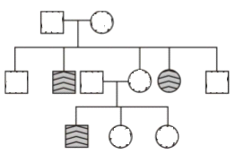
Question: From NCERT NEET [Difficult level:Easy]
Study the pedigree chart of a family showing the inheritance of myotonic dystrophy.
The trait under study is
(1) dominant X-linked
(2) recessive X-linked
(3) autosomal dominant
(4) recessive Y-linked.
Answer ▽
(3) autosomal dominant
⬆️Prev____@organised notes_____Next⬇️
Question: From NCERT NEET [Difficult level:Easy]
One of the genes present exclusively on the X-chromosome in humans is concerned with
(1) baldness
(2) red-green colour blindness
(3) facial hair/moustaches in males
(d) night blindness
Answer ▽
(2) red-green colour blindness
Colour Blidness :
either red or green cone of eye resulting in failure to discriminate between
red and green colour. This defect is due to mutation in certain genes
present in the X chromosome. It occurs in about 8 per cent of males and
only about 0.4 per cent of females. This is because the genes that lead to
red-green colour blindness are on the X chromosome.
one X chromosome and females have two. The son of a woman
not herself colour blind because the gene is recessive. That means that its
effect is suppressed by her matching dominant normal gene. A daughter
will not normally be colour blind, unless her mother is a carrier and her
father is colour blind.
Haemophilia :
transmission from unaffected carrier female to some of the male progeny
has been widely studied. In this disease, a single protein that is a part of
the cascade of proteins involved in the clotting of blood is affected. Due to
this, in an affected individual a simple cut will result in non-stop bleeding.
The heterozygous female (carrier) for haemophilia may transmit the disease
to sons. The possibility of a female becoming a haemophilic is extremely
rare because mother of such a female has to be at least carrier and the
father should be haemophilic (unviable in the later stage of life). The family
pedigree of Queen Victoria shows a number of haemophilic descendents
as she was a carrier of the disease.
Sickle-cell anaemia :
be transmitted from parents to the offspring when both the partners are
carrier for the gene (or heterozygous). The disease is controlled by a single
pair of allele, HbA and HbS. Out of the three possible genotypes only
homozygous individuals for HbS (HbSHbS) show the diseased phenotype.
Heterozygous (HbAHbS) individuals appear apparently unaffected but they
are carrier of the disease as there is 50 per cent probability of transmission
of the mutant gene to the progeny, thus exhibiting sickle-cell trait
(Figure 5.15). The defect is caused by the substitution of Glutamic acid (Glu) by Valine (Val) at the sixth position of the beta globin chain of the
haemoglobin molecule. The substitution of amino acid in the globin
protein results due to the single base substitution at the sixth codon of
the beta globin gene from GAG to GUG. The mutant haemoglobin molecule
undergoes polymerisation under low oxygen tension causing the change
in the shape of the RBC from biconcave disc to elongated sickle like
structure (Figure 5.15).
Phenylketonuria :
the autosomal recessive trait. The affected individual lacks an enzyme
that converts the amino acid phenylalanine into tyrosine. As a result of
this phenylalanine is accumulated and converted into phenylpyruvic acid
and other derivatives. Accumulation of these in brain results in mental
retardation. These are also excreted through urine because of its poor
absorption by kidney.
Thalassemia :
transmitted from parents to the offspring when both the partners are
unaffected carrier for the gene (or heterozygous). The defect could be due
to either mutation or deletion which ultimately results in reduced rate of
synthesis of one of the globin chains (a and b chains) that make up
haemoglobin. This causes the formation of abnormal haemoglobin
molecules resulting into anaemia which is characteristic of the disease.
Thalassemia can be classified according to which chain of the haemoglobin
molecule is affected. In a Thalassemia, production of a globin chain is
affected while in b Thalassemia, production of b globin chain is affected.
a Thalassemia is controlled by two closely linked genes HBA1 and HBA2
on chromosome 16 of each parent and it is observed due to mutation or
deletion of one or more of the four genes. The more genes affected, the
less alpha globin molecules produced. While b Thalassemia is controlled
by a single gene HBB on chromosome 11 of each parent and occurs due
to mutation of one or both the genes. Thalassemia differs from sickle-cell
anaemia in that the former is a quantitative problem of synthesising too
few globin molecules while the latter is a qualitative problem of
synthesising an incorrectly functioning globin.
⬆️Prev____@organised notes_____Next⬇️
Question: From NCERT NEET [Difficult level:Easy]
Which of the following occurs due to monosomy of sex chromosome?
(1) Down's syndrome
(2) Turner's syndrome
(3) Haemophilia
(4) Sickle cell anaemia
Answer ▽
(2) Turner's syndrome
Down’s Syndrome :
The cause of this genetic disorder
is the presence of an additional copy of the
chromosome number 21 (trisomy of 21). This disorder
was first described by Langdon Down (1866). The
affected individual is short statured with small round
head, furrowed tongue and partially open mouth
(Figure 5.16). Palm is broad with characteristic palm
crease. Physical, psychomotor and mental
development is retarded.
Klinefelter’s Syndrome :
This genetic disorder is also
caused due to the presence of an additional copy of Xchromosome
resulting into a karyotype of 47, XXY.
Such an individual has overall masculine development,
however, the feminine development (development
of breast, i.e., Gynaecomastia) is also expressed
(Figure 5.17 a). Such individuals are sterile.
Turner’s Syndrome :
Such a disorder is caused due
to the absence of one of the X chromosomes, i.e., 45 with X0, Such females
are sterile as ovaries are rudimentary besides other features including
lack of other secondary sexual characters .
⬆️Prev____@organised notes_____Next⬇️
Question: From NCERT NEET [Difficult level:Easy] AIIMS - 2007
XO-chromosomal abnormality in human beings causes
(1) Turner's syndrome
(2) Down's syndrome
(3) Kilnefelter's syndrome
(4) none of these.
Answer ▽
(1) Turner's syndrome
Down’s Syndrome :
The cause of this genetic disorder
is the presence of an additional copy of the
chromosome number 21 (trisomy of 21). This disorder
was first described by Langdon Down (1866). The
affected individual is short statured with small round
head, furrowed tongue and partially open mouth
(Figure 5.16). Palm is broad with characteristic palm
crease. Physical, psychomotor and mental
development is retarded.
Klinefelter’s Syndrome :
This genetic disorder is also
caused due to the presence of an additional copy of Xchromosome
resulting into a karyotype of 47, XXY.
Such an individual has overall masculine development,
however, the feminine development (development
of breast, i.e., Gynaecomastia) is also expressed
(Figure 5.17 a). Such individuals are sterile.
Turner’s Syndrome :
Such a disorder is caused due
to the absence of one of the X chromosomes, i.e., 45 with X0, Such females
are sterile as ovaries are rudimentary besides other features including
lack of other secondary sexual characters .
⬆️Prev____@organised notes_____Next⬇️
Question: From NCERT NEET [Difficult level:Easy]
Which one of the following pairs of features is a good example of polygenic inheritance?
(1) Human height and skin colour
(2) ABO blood group in humans and flower colour of Mirabilis jalapa
(3) Hair pigment of mouse and tongue rolling in humans
(4) Human eye colour and sickle cell anaemia
Answer ▽
(1) Human height and skin colour
POLYGENIC INHERITANCE
Mendel’s studies mainly described those traits that have distinct alternate
forms such as flower colour which are either purple or white. But if you
look around you will find that there are many traits which are not so
distinct in their occurrence and are spread across a gradient. For example,
in humans we don’t just have tall or short people as two distinct
alternatives but a whole range of possible heights. Such traits are generally
controlled by three or more genes and are thus called as polygenic traits.
Besides the involvement of multiple genes polygenic inheritance also takes
into account the influence of environment. Human skin colour is another
classic example for this. In a polygenic trait the phenotype reflects the
contribution of each allele, i.e., the effect of each allele is additive. To
understand this better let us assume that three genes A, B, C control skin
colour in human with the dominant forms A, B and C responsible for
dark skin colour and the recessive forms a, b and c for light skin colour.
The genotype with all the dominant alleles (AABBCC) will have the darkest
skin colour and that with all the recessive alleles (aabbcc) will have the
lightest skin colour. As expected the genotype with three dominant alleles
and three recessive alleles will have an intermediate skin colour. In this
manner the number of each type of alleles in the genotype would determine
the darkness or lightness of the skin in an individual.
⬆️Prev____@organised notes_____Next⬇️
Question: From NCERT NEET [Difficult level:Easy]
Sickle cell anaemia is example of
(1) sex-linked inheritance
(2) deficiency disease
(3) autosomal heritable disease
(4) infectious disease
Answer ▽
(3) autosomal heritable disease
Colour Blidness :
either red or green cone of eye resulting in failure to discriminate between
red and green colour. This defect is due to mutation in certain genes
present in the X chromosome. It occurs in about 8 per cent of males and
only about 0.4 per cent of females. This is because the genes that lead to
red-green colour blindness are on the X chromosome. Males have only
one X chromosome and females have two. The son of a woman
not herself colour blind because the gene is recessive. That means that its
effect is suppressed by her matching dominant normal gene. A daughter
will not normally be colour blind, unless her mother is a carrier and her
father is colour blind.
Haemophilia :
transmission from unaffected carrier female to some of the male progeny
has been widely studied. In this disease, a single protein that is a part of
the cascade of proteins involved in the clotting of blood is affected. Due to
this, in an affected individual a simple cut will result in non-stop bleeding.
The heterozygous female (carrier) for haemophilia may transmit the disease
to sons. The possibility of a female becoming a haemophilic is extremely
rare because mother of such a female has to be at least carrier and the
father should be haemophilic (unviable in the later stage of life). The family
pedigree of Queen Victoria shows a number of haemophilic descendents
as she was a carrier of the disease.
Sickle-cell anaemia :
be transmitted from parents to the offspring when both the partners are
carrier for the gene (or heterozygous). The disease is controlled by a single
pair of allele, HbA and HbS. Out of the three possible genotypes only
homozygous individuals for HbS (HbSHbS) show the diseased phenotype.
Heterozygous (HbAHbS) individuals appear apparently unaffected but they
are carrier of the disease as there is 50 per cent probability of transmission
of the mutant gene to the progeny, thus exhibiting sickle-cell trait
(Figure 5.15). The defect is caused by the substitution of Glutamic acid (Glu) by Valine (Val) at the sixth position of the beta globin chain of the
haemoglobin molecule. The substitution of amino acid in the globin
protein results due to the single base substitution at the sixth codon of
the beta globin gene from GAG to GUG. The mutant haemoglobin molecule
undergoes polymerisation under low oxygen tension causing the change
in the shape of the RBC from biconcave disc to elongated sickle like
structure (Figure 5.15).
Phenylketonuria :
the autosomal recessive trait. The affected individual lacks an enzyme
that converts the amino acid phenylalanine into tyrosine. As a result of
this phenylalanine is accumulated and converted into phenylpyruvic acid
and other derivatives. Accumulation of these in brain results in mental
retardation. These are also excreted through urine because of its poor
absorption by kidney.
Thalassemia :
transmitted from parents to the offspring when both the partners are
unaffected carrier for the gene (or heterozygous). The defect could be due
to either mutation or deletion which ultimately results in reduced rate of
synthesis of one of the globin chains (a and b chains) that make up
haemoglobin. This causes the formation of abnormal haemoglobin
molecules resulting into anaemia which is characteristic of the disease.
Thalassemia can be classified according to which chain of the haemoglobin
molecule is affected. In a Thalassemia, production of a globin chain is
affected while in b Thalassemia, production of b globin chain is affected.
a Thalassemia is controlled by two closely linked genes HBA1 and HBA2
on chromosome 16 of each parent and it is observed due to mutation or
deletion of one or more of the four genes. The more genes affected, the
less alpha globin molecules produced. While b Thalassemia is controlled
by a single gene HBB on chromosome 11 of each parent and occurs due
to mutation of one or both the genes. Thalassemia differs from sickle-cell
anaemia in that the former is a quantitative problem of synthesising too
few globin molecules while the latter is a qualitative problem of
synthesising an incorrectly functioning globin.
⬆️Prev____@organised notes_____Next⬇️
Question: From NCERT NEET [Difficult level:Easy]
How many pairs of contrasting traits studied by Mendel were related to pod?
(1) 1
(2) 2
(3) 3
(4) 4
Answer ▽
(2) 2
⬆️Prev____@organised notes_____Next⬇️
Question: From NCERT NEET [Difficult level:Easy]
“Filial 1 progeny” refers to
(1) First pure breed
(2) First offspring
(3) First hybrid generation
(4) Second hybrid generation
Answer ▽
(3) First hybrid generation
INHERITANCE OF ONE GENE
Let us take the example of one such
hybridisation experiment carried out by
Mendel where he crossed tall and dwarf pea
plants to study the inheritance of one gene
(Figure 5.2). He collected the seeds produced
as a result of this cross and grew them to
generate plants of the first hybrid generation.
This generation is also called the Filial1
progeny or the F1. Mendel observed that all
the F1 progeny plants were tall, like one of
its parents; none were dwarf (Figure 5.3). He
made similar observations for the other pairs
of traits – he found that the F1 always
resembled either one of the parents, and that
the trait of the other parent was not seen in
them.
Mendel then self-pollinated the tall F1
plants and to his surprise found that in the
Filial2 generation some of the offspring were
‘dwarf ’; the character that was not seen in
the F1 generation was now expressed. The
proportion of plants that were dwarf were
1/4th of the F2 plants while 3/4th of the F2 plants were tall. The tall and
dwarf traits were identical to their parental type and did not show any
blending, that is all the offspring were either tall or dwarf, none were of inbetween
height (Figure 5.3).
Similar results were obtained with the other traits that he studied:
only one of the parental traits was expressed in the F1 generation while at
the F2 stage both the traits were expressed in the proportion 3:1. The
contrasting traits did not show any blending at either F1 or F2 stage.
⬆️Prev____@organised notes_____Next⬇️
Question: From NCERT NEET [Difficult level:Easy]
Gregor Mendel is known for
(1) Species concept
(2) Hybridisation experiments
(3) Discovery of chromosomes
(4) Experiments on maize plant
Answer ▽
(2) Hybridisation experiments
INHERITANCE OF ONE GENE
Let us take the example of one such
hybridisation experiment carried out by
Mendel where he crossed tall and dwarf pea
plants to study the inheritance of one gene
(Figure 5.2). He collected the seeds produced
as a result of this cross and grew them to
generate plants of the first hybrid generation.
This generation is also called the Filial1
progeny or the F1. Mendel observed that all
the F1 progeny plants were tall, like one of
its parents; none were dwarf (Figure 5.3). He
made similar observations for the other pairs
of traits – he found that the F1 always
resembled either one of the parents, and that
the trait of the other parent was not seen in
them.
Mendel then self-pollinated the tall F1
plants and to his surprise found that in the
Filial2 generation some of the offspring were
‘dwarf ’; the character that was not seen in
the F1 generation was now expressed. The
proportion of plants that were dwarf were
1/4th of the F2 plants while 3/4th of the F2 plants were tall. The tall and
dwarf traits were identical to their parental type and did not show any
blending, that is all the offspring were either tall or dwarf, none were of inbetween
height (Figure 5.3).
Similar results were obtained with the other traits that he studied:
only one of the parental traits was expressed in the F1 generation while at
the F2 stage both the traits were expressed in the proportion 3:1. The
contrasting traits did not show any blending at either F1 or F2 stage.
⬆️Prev____@organised notes_____Next⬇️
Question: From NCERT NEET [Difficult level:Easy]
A cross between tall pea plant and its recessive parent is known as
(1) Back cross
(2) Test cross
(3) Recessive cross
(4) Dominant cross
Answer ▽
(2) Test cross
whether a tall plant from F1
or F2 has TT or Tt composition, cannot be predicted. Therefore, to determine
the genotype of a tall plant at F2, Mendel crossed the tall plant from F2
with a dwarf plant. This he called a test cross. In a typical test cross an
organism (pea plants here) showing a dominant phenotype (and whose
genotype is to be determined) is crossed with the recessive parent instead
of self-crossing. The progenies of such a cross can easily be analysed to
predict the genotype of the test organism
⬆️Prev____@organised notes_____Next⬇️
Question: From NCERT NEET [Difficult level:Easy]
Who was the first to use statistical analysis and mathematical logics for solving biological problems?
1) Francis crick
(2) Gregor Mendel
(3) Hugo de vries
(4) W. Bateson
Answer ▽
(2) Gregor Mendel
-Gregor Mendel, conducted
hybridisation experiments on garden peas for
seven years (1856-1863) and proposed the
laws of inheritance in living organisms. During
Mendel’s investigations into inheritance
patterns it was for the first time that statistical
analysis and mathematical logic were applied
to problems in biology. His experiments had a
large sampling size, which gave greater
credibility to the data that he collected. Also,
the confirmation of his inferences from
experiments on successive generations of his
test plants, proved that his results pointed to
general rules of inheritance rather than being
unsubstantiated ideas. Mendel investigated
characters in the garden pea plant that were
manifested as two opposing traits, e.g., tall or
dwarf plants, yellow or green seeds. This
allowed him to set up a basic framework of
rules governing inheritance, which was
expanded on by later scientists to account for
all the diverse natural observations and the
complexity inherent in them.
⬆️Prev____@organised notes_____Next⬇️
Question: From NCERT NEET [Difficult level:Easy]
Trait expressed in F1 generation is
(1) Parental
(2) Recessive
(3) Dominant
(4) Recombinant
Answer ▽
(3) Dominant
Mendel observed that all
the F1 progeny plants were tall, like one of
its parents; none were dwarf (Figure 5.3). He
made similar observations for the other pairs
of traits – he found that the F1 always
resembled either one of the parents, and that
the trait of the other parent was not seen in
them.
The law of dominance is used to explain the expression of only one of
the parental characters in a monohybrid cross in the F1 and the expression
of both in the F2. It also explains the proportion of 3:1 obtained at the F2.
⬆️Prev____@organised notes_____Next⬇️
Question: From NCERT NEET [Difficult level:Easy]
Genotypic and phenotypic ratios of monohybrid cross are
(1) 3:1 and 1:2:1
(2) 1:2:1 and 3:1
(3) 1:2:1 and 1:2:1
(4) 3:1 and 3:1
Answer ▽
(2) 1:2:1 and 3:1
⬆️Prev____@organised notes_____Next⬇️
Question: From NCERT NEET [Difficult level:Easy]
Incomplete dominance is present in
(1) Snapdragon flower
(2) Dog flower
(3) Antirrhinum flower
(4) All of the above
Answer ▽
(4) All of the above
Incomplete Dominance
When experiments on peas were repeated using other
traits in other plants, it was found that sometimes
the F1 had a phenotype that did not resemble either
of the two parents and was in between the two. The
inheritance of flower colour in the dog flower
(snapdragon or Antirrhinum sp.) is a good example
to understand incomplete dominance. In a cross
between true-breeding red-flowered (RR) and truebreeding
white-flowered plants (rr), the F1 (Rr) was
pink (Figure 5.6). When the F1 was self-pollinated
the F2 resulted in the following ratio 1 (RR) Red: 2
(Rr) Pink: 1 (rr) White. Here the genotype ratios were
exactly as we would expect in any mendelian
monohybrid cross, but the phenotype ratios had
changed from the 3:1 dominant : recessive ratio.
What happened was that R was not completely
dominant over r and this made it possible to
distinguish Rr as pink from RR (red) and rr (white) .
Explanation of the concept of dominance:
What exactly is dominance? Why are some alleles
dominant and some recessive? To tackle these
questions, we must understand what a gene does.
Every gene, as you know by now, contains the
information to express a particular trait. In a
diploid organism, there are two copies of each
gene, i.e., as a pair of alleles. Now, these two alleles
need not always be identical, as in a heterozygote.
One of them may be different due to some changes
that it has undergone (about which you will read
further on, and in the next chapter) which modifies
the information that particular allele contains.
Let’s take an example of a gene that contains
the information for producing an enzyme. Now
there are two copies of this gene, the two allelic
forms. Let us assume (as is more common) that
the normal allele produces the normal enzyme
that is needed for the transformation of a
substrate S. Theoretically, the modified allele could be responsible for
production of –
(i) the normal/less efficient enzyme, or
(ii) a non-functional enzyme, or
(iii) no enzyme at all
⬆️Prev____@organised notes_____Next⬇️
Question: From NCERT NEET [Difficult level:Easy]
In human ABO blood grouping possible number of genotypes is
(1) 3
(2) 4
(3) 6
(4) 9
Answer ▽
(3) 6
ABO blood groups are controlled by
the gene I. The plasma membrane of the red blood cells has sugar polymers
that protrude from its surface and the kind of sugar is controlled by the
gene. The gene (I) has three alleles IA, IB and i. The alleles IA and IB produce
a slightly different form of the sugar while allele i does not produce any
sugar. Because humans are diploid organisms, each person possesses
any two of the three I gene alleles. IA and IB are completely dominant over
i, in other words when IA and i are present only IA expresses (because i
does not produce any sugar), and when IB and i are present IB expresses.
But when IA and IB are present together they both express their own types
of sugars: this is because of co-dominance. Hence red blood cells have
both A and B types of sugars. Since there are three different alleles, there
are six different combinations of these three alleles that are possible, and
therefore, a total of six different genotypes of the human ABO blood types
⬆️Prev____@organised notes_____End