

.The human skull has 22 bones with ..................cranial bones and ............................ facial bones
1. 10,12
2. 14,8
3. 12,10
4. 8,14
.
.
Answer ▽ ✅Verified
4. 8,14
SKELETAL SYSTEM
Skeletal system consists of a framework of bones and a few cartilages.This system has a significant role in movement shown by the body.
Imagine chewing food without jaw bones and walking around without
the limb bones. Bone and cartilage are specialised connective tissues.
The former has a very hard matrix due to calcium salts in it and the latter
has slightly pliable matrix due to chondroitin salts. In human beings,
this system is made up of 206 bones and a few cartilages. It is grouped
into two principal divisions – the axial and the appendicular skeleton.
Axial skeleton comprises 80 bones distributed along the main axis
of the body. The skull, vertebral column, sternum and ribs constitute
axial skeleton. The skull is composed of two sets of bones –
⬆️Prev____@organised notes_____Next⬇️
.Which of the following has ATPase activity?
1. Actin 2. Tropomyosin
3. Head of myosin 4. Troponin
.
.
Answer ▽ ✅Verified
3. Head of myosin
⬆️Prev____@organised notes_____Next⬇️
.The ATPase activity of the myosin head is dependent on:
1. Magnesium ions
2. Manganese ions
3. Calcium ions
4. Ferric ions
.
.
Answer ▽ ✅Verified
3. Calcium ions
system (CNS) via a motor neuron. A motor neuron alongwith the muscle
fibres connected to it constitute a motor unit. The junction between a
motor neuron and the sarcolemma of the muscle fibre is called the
neuromuscular junction or motor-end plate. A neural signal reaching
this junction releases a neurotransmitter (Acetyl choline) which generates
an action potential in the sarcolemma. This spreads through the muscle
fibre and causes the release of calcium ions into the sarcoplasm. Increase
in Ca++ level leads to the binding of calcium with a subunit of troponin on
actin filaments and thereby remove the masking of active sites for myosin.
Utilising the energy from ATP hydrolysis, the myosin head now binds to
the exposed active sites on actin to form a cross bridge
⬆️Prev____@organised notes_____Next⬇️
.During muscle contraction, the length of all the following gets reduced except:
1. H-Zone
2. A-Band
3. I-Band
4. Sacromere
.
.
Answer ▽ ✅Verified
2. A-Band
Muscle contraction is initiated by a signal sent by the central nervous
system (CNS) via a motor neuron. A motor neuron alongwith the muscle
fibres connected to it constitute a motor unit. The junction between a
motor neuron and the sarcolemma of the muscle fibre is called the
neuromuscular junction or motor-end plate. A neural signal reaching
this junction releases a neurotransmitter (Acetyl choline) which generates
an action potential in the sarcolemma. This spreads through the muscle
fibre and causes the release of calcium ions into the sarcoplasm. Increase
in Ca++ level leads to the binding of calcium with a subunit of troponin on
actin filaments and thereby remove the masking of active sites for myosin.
Utilising the energy from ATP hydrolysis, the myosin head now binds to
‘Z’ line attached to these actins are also pulled inwards thereby causing a
shortening of the sarcomere, i.e., contraction. It is clear from the above
steps, that during shortening of the muscle, i.e., contraction, the ‘I’ bands
get reduced, whereas the ‘A’ bands retain the length.

⬆️Prev____@organised notes_____Next⬇️
.What is true about the white muscle fibers?
1. Myoglobin content is high
2. They have a large number of mitochondria
3. They depend on anaerobic process for energy
4. They are adapted for slow sustained activities
.
.
Answer ▽ ✅Verified
3. They depend on anaerobic process for energy
Muscle contains a red coloured oxygen storing
pigment called myoglobin. Myoglobin content is high in some of the
muscles which gives a reddish appearance. Such muscles are called the
Red fibres. These muscles also contain plenty of mitochondria which can
utilise the large amount of oxygen stored in them for ATP production.
These muscles, therefore, can also be called aerobic muscles. On the
other hand, some of the muscles possess very less quantity of myoglobin
and therefore, appear pale or whitish. These are the White fibres. Number
of mitochondria are also few in them, but the amount of sarcoplasmic
reticulum is high. They depend on anaerobic process for energy.
Why do white muscle fibres have more sarcoplasmic reticulum?
White muscle fibres are designed for quick movements (like the muscles in your hands & for moving your eyes). Also they are used for high energy activity for short periods of time. Due to this they need more sarcoplasmic reticulum so that they are better equipped for quick release and re-uptake of calcium ions.
⬆️Prev____@organised notes_____Next⬇️
.Scapula is a large triangular flat bone situated in the dorsal part of the thorax between:
1. the second and fifth ribs
2. the second and seventh ribs
3. the third and sixth ribs
4. the third and eighth ribs
.
.
Answer ▽ ✅Verified
2. the second and seventh ribs
the articulation of the upper and the lower limbs
respectively with the axial skeleton. Each
scapula . Scapula is a large
triangular flat bone situated in the dorsal part
of the thorax between the second and the
seventh ribs. The dorsal, flat, triangular body
of scapula has a slightly elevated ridge called
the spine which projects as a flat, expanded
process called the acromion. The clavicle
articulates with this. Below the acromion is a
depression called the glenoid cavity which
articulates with the head of the humerus to
form the shoulder joint. Each clavicle is a long
slender bone with two curvatures. This bone
is commonly called the collar bone.
Pelvic girdle consists of two coxal bones
. Each coxal bone is formed by
the fusion of three bones – ilium, ischium and
pubis. At the point of fusion of the above bones
is a cavity called acetabulum to which the thigh
bone articulates. The two halves of the pelvic
girdle meet ventrally to form the pubic
symphysis containing fibrous cartilage.
⬆️Prev____@organised notes_____Next⬇️
.Match each item in Column I with one item in Column II and chose your answer from the codes given below:
Column I
Column II
I. Acetabulum
II. Glenoid Cavity
III. Vertebrosternal ribs
IV. Vertebrochondral ribs
1. Pectoral girdie
2. Pelvic girdie
3. 7 pairs
4. 3 pairs
Codes
I II III IV
(1) 1 2 3 4
(2) 2 1 4 3
(3) 2 1 3 4
(4) 1 2 4 3
.
.
Answer ▽ ✅Verified
(3) 2 1 3 4
the articulation of the upper and the lower limbs
respectively with the axial skeleton. Each
scapula . Scapula is a large
triangular flat bone situated in the dorsal part
of the thorax between the second and the
seventh ribs. The dorsal, flat, triangular body
of scapula has a slightly elevated ridge called
the spine which projects as a flat, expanded
process called the acromion. The clavicle
articulates with this. Below the acromion is a
depression called the glenoid cavity which
articulates with the head of the humerus to
form the shoulder joint. Each clavicle is a long
slender bone with two curvatures. This bone
is commonly called the collar bone.
Pelvic girdle consists of two coxal bones
. Each coxal bone is formed by
the fusion of three bones – ilium, ischium and
pubis. At the point of fusion of the above bones
is a cavity called acetabulum to which the thigh
bone articulates. The two halves of the pelvic
girdle meet ventrally to form the pubic
symphysis containing fibrous cartilage.
⬆️Prev____@organised notes_____Next⬇️
.The pivot joint between atlas and axis is a type of
(1) fibrous joint
(2) cartilaginous joint
(3) synovial joint
(4) saddle joint
.
.
Answer ▽ ✅Verified
(3) synovial joint
JOINTS
involving the bony parts of the body.
⬆️Prev____@organised notes_____Next⬇️
.Select the correct matching of the type of the joint with the example in human skeletal
system
Types of joint Example
1. Cartilaginous joint Between frontal and parietal
2. Pivot joint Between third and fourth Cervical vertebrae
3. Hinge joint Between humerus and pectoral girdle
4. Gliding joint Between carpals
.
.
Answer ▽ ✅Verified
4. Gliding joint Between carpals
JOINTS
involving the bony parts of the body.
⬆️Prev____@organised notes_____Next⬇️
.The characteristics and an example of a synovial joint in humans is
| Characteristics | Examples |
1. | Fluid cartilage between two bones, limited movements | Knee joints |
2. | Fluid filled between two joints, provides cushion | Skull bones |
3. | Fluid filled synovial cavity between two bones | Joint between atlas and axis |
4. | Lymph filled between two bones, limited movement | Gliding joint between carpals |
.
.
Answer ▽ ✅Verified
4. | Lymph filled between two bones, limited movement | Gliding joint between carpals |
.JOINTS
involving the bony parts of the body.
Saliva is mainly produced by three pairs of
salivary glands, the parotids (cheek), the submaxillary/
sub-mandibular (lower jaw) and the
sub- linguals (below the tongue).
⬆️Prev____@organised notes_____Next⬇️
.Given diagram shows bone of the left human hindlimb as seen from front. It has certain mistakes in labeling. Two of the wrongly labelled bones are
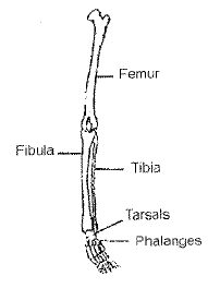
(1) tibia and tarsals
(2) femur and fibula
(3) fibula and phalanges
(4) tarsals and femur
.
.
Answer ▽ ✅Verified
(3) fibula and phalanges

⬆️Prev____@organised notes_____Next⬇️
.Skeletal muscles appear striated due to presence of two characteristic protiens in alternating dark and light bands. Which of the following is a correct match of the protien with its light refractive property and colour?
Protien Colour Property
(1) Myosin Light Anisotropic
(2) Actin Dark Anisotropic
(3) Myosin Dark Isotropic
(4) Actin Light Isotropic
.
.
Answer ▽ ✅Verified
(4) Actin Light Isotropic
A characteristic feature of the muscle fibre is the presence of a large number
of parallelly arranged filaments in the sarcoplasm called myofilaments or
myofibrils. Each myofibril has alternate dark and light bands on it. A
detailed study of the myofibril has established that the striated appearance
is due to the distribution pattern of two important proteins – Actin and
Myosin. The light bands contain actin and is called I-band or Isotropic
band, whereas the dark band called ‘A’ or Anisotropic band
⬆️Prev____@organised notes_____Next⬇️
.During muscular contraction, which of the following events occur?
(i) H-zone disappears
(ii) A band widens
(iii) I band shortens
(iv) Width of A band is unaffected
(v) M line and Z line come closer
(1) (i),(iii),(iv) and (v)
(2) (i),(ii) and (v)
(3) (ii),(iv) and (v)
(4) (i),(ii) and (iii).
.
.
Answer ▽ ✅Verified
(1) (i),(iii),(iv) and (v)

⬆️Prev____@organised notes_____Next⬇️
.Which of the following is true for the labelled parts in the figure below?
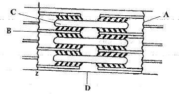
(1) A - Z-line - located at centre of I - band
(2) B - Thin filament - occurs in A-band only
(3) C - Thick filament - confined to I-band
(4) D - H-zone - located at centre of M-line
.
.
Answer ▽ ✅Verified
(1) A - Z-line - located at centre of I - band
⬆️Prev____@organised notes_____Next⬇️
.Pick out the correct match
(1) Sternum - 14
(2) Pelvis - 3
(3) Ribs - 20
(4) Face - 5
.
.
Answer ▽ ✅Verified
(2) Pelvis - 3
the articulation of the upper and the lower limbs
respectively with the axial skeleton. Each
scapula . Scapula is a large
triangular flat bone situated in the dorsal part
of the thorax between the second and the
seventh ribs. The dorsal, flat, triangular body
of scapula has a slightly elevated ridge called
the spine which projects as a flat, expanded
process called the acromion. The clavicle
articulates with this. Below the acromion is a
depression called the glenoid cavity which
articulates with the head of the humerus to
form the shoulder joint. Each clavicle is a long
slender bone with two curvatures. This bone
is commonly called the collar bone.
Pelvic girdle consists of two coxal bones
. Each coxal bone is formed by
the fusion of three bones – ilium, ischium and
pubis. At the point of fusion of the above bones
is a cavity called acetabulum to which the thigh
bone articulates. The two halves of the pelvic
girdle meet ventrally to form the pubic
symphysis containing fibrous cartilage.
⬆️Prev____@organised notes_____Next⬇️
.Sarcomere is the area between:
(1) 2 H-zones
(2) 2 Z-lines
(3) 2 M-lines
(4) 2 A-bands
.
.
Answer ▽ ✅Verified
(2) 2 Z-lines
Actin
filaments are thinner as compared to the myosin filaments, hence are
commonly called thin and thick filaments respectively. In the centre of
each ‘I’ band is an elastic fibre called ‘Z’ line which bisects it. The thin
filaments are firmly attached to the ‘Z’ line. The thick filaments in the
‘A’ band are also held together in the middle of this band by a thin fibrous
membrane called ‘M’ line. The ‘A’ and ‘I’ bands are arranged alternately
throughout the length of the myofibrils. The portion of the myofibril
between two successive ‘Z’ lines is considered as the functional unit of
contraction and is called a sarcomere
⬆️Prev____@organised notes_____Next⬇️
.Go through the following diagram describing muscle contraction:
Now identify A to E:
(1) A - Cross bridge, B - Cross bridge formation, C - Breaking of cross bridge, D - Sliding (rotation), E - ATP
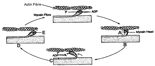
(2) A - Cross bridge, B - Cross bridge formation, C - Sliding / rotation, D - Breaking of cross bridge, E - ATP
(3) A - Cross bridge, B - Breaking of Cross bridge, C - sliding / rotation, D - Cross bridge formation, E - AMP
(4) A - Cross bridge, B - Cross bridge formation, C - Sliding / rotation, D - ADP, E - Breaking of cross bridge
.
.
Answer ▽ ✅Verified
(2) A - Cross bridge, B - Cross bridge formation, C - Sliding / rotation, D - Breaking of cross bridge, E - ATP

⬆️Prev____@organised notes_____Next⬇️
.I. Number of mitochondria less.
II. Number of mitochondria more
III. Sarcoplasmic reticulum is abundant
IV. Myoglobin content high
V. Sarcoplasmic reticulum moderate
VI. Aerobic muscles
VII. Depend on anaerobic respiration for energy
VIII. Less myoglobin content
A. Red muscles B. White muscles
Identify above (I to VIII) traits as characteristic of A and B types of muscles:
(1) A - I, III, VII, VIII; B - II, IV, V, VI
(2) A - II, IV, V, VI; B - I, III, VII, VIII
(3) A - I, III, IV, VII; B - II, V, VI, VIII
(4) A - II, V, VI, VIII; B - I, III, IV, VII
.
.
Answer ▽ ✅Verified
(2) A - II, IV, V, VI; B - I, III, VII, VIII
Muscle contains a red coloured oxygen storing
pigment called myoglobin. Myoglobin content is high in some of the
muscles which gives a reddish appearance. Such muscles are called the
Red fibres. These muscles also contain plenty of mitochondria which can
utilise the large amount of oxygen stored in them for ATP production.
These muscles, therefore, can also be called aerobic muscles. On the
other hand, some of the muscles possess very less quantity of myoglobin
and therefore, appear pale or whitish. These are the White fibres. Number
of mitochondria are also few in them, but the amount of sarcoplasmic
reticulum is high. They depend on anaerobic process for energy.
Why do white muscle fibres have more sarcoplasmic reticulum?
White muscle fibres are designed for quick movements (like the muscles in your hands & for moving your eyes). Also they are used for high energy activity for short periods of time. Due to this they need more sarcoplasmic reticulum so that they are better equipped for quick release and re-uptake of calcium ions.
⬆️Prev____@organised notes_____Next⬇️
.Match Column-I with Column-II:
Column-I Column-II (Number of bones)
A. Cranium / Brainbox I. 22
B. Skull (Cranial and facial bones) II. 8
C. Face III. 14
D. Hind limb IV. 12 pairs
E. Ribs V. 30
(1) A - I, B - II, C - III, D - V, E - IV
(2) A - II, B - I, C - III, D - V, E - IV
(3) A - I, B - II, C - III, D - IV, E - V
(4) A - V, B - IV, C - III, D - II, A - I
.
.
Answer ▽ ✅Verified
(2) A - II, B - I, C - III, D - V, E - IV
SKELETAL SYSTEM
Skeletal system consists of a framework of bones and a few cartilages.This system has a significant role in movement shown by the body.
Imagine chewing food without jaw bones and walking around without
the limb bones. Bone and cartilage are specialised connective tissues.
The former has a very hard matrix due to calcium salts in it and the latter
has slightly pliable matrix due to chondroitin salts. In human beings,
this system is made up of 206 bones and a few cartilages. It is grouped
into two principal divisions – the axial and the appendicular skeleton.
Axial skeleton comprises 80 bones distributed along the main axis
of the body. The skull, vertebral column, sternum and ribs constitute
axial skeleton. The skull is composed of two sets of bones –
⬆️Prev____@organised notes_____Next⬇️
.Human adult vertebral formula is:
(1) C4 T8 L4 S8 C8
(2) C7 T8 L5 S6 C7
(3) C7 T112 L2 S1 C2
(4) C7 T12 L5 S1 C1
.
.
Answer ▽ ✅Verified
(4) C7 T12 L5 S1 C1
⬆️Prev____@organised notes_____Next⬇️
.Match the Column-I with Column-II:
Column-I Column-II
A. True ribs I. 3 pairs
B. False ribs II. 2 pairs
C. Floating ribs III. 7 pairs
(1) A - I, B - II, C - III
(2) A - III, B - I, C - II
(3) A - III, B - II, C - I
(4) A - II, B - I, C - III
.
.
Answer ▽ ✅Verified
(2) A - III, B - I, C - II
⬆️Prev____@organised notes_____Next⬇️
.Number of bone in each upper limb is:
(1) 1, 1, 1
(2) 8, 5, 14
(3) 2, 2, 2, 16, 10, 28
(4) 1, 1, 1, 8, 5, 14
.
.
Answer ▽ ✅Verified
(4) 1, 1, 1, 8, 5, 14
⬆️Prev____@organised notes_____Next⬇️
.An acromion process is characteristically found in:
(1) Pelvic girdle of mammals
(2) Pectoral girdle of mammals
(3) Skull bone
(4) Vertebrae of mammals
.
.
Answer ▽ ✅Verified
(2) Pectoral girdle of mammals
⬆️Prev____@organised notes_____Next⬇️
.Acetabulum occurs in:
(1) Cranium
(2) Pectoral girdle
(3) Pelvic girdle
(4) Vertebrae
.
.
Answer ▽ ✅Verified
(3) Pelvic girdle
⬆️Prev____@organised notes_____Next⬇️
.Each coxal bone is formed by the fusion of 3 bones name .as:
(a) Ileum, ischium and pubis
b. Ilium, ischium and pubis
(c) Ilium, ischium and clavicle
(d) Coracoid, ischium and pubis
.
.
Answer ▽ ✅Verified
b. Ilium, ischium and pubis
⬆️Prev____@organised notes_____Next⬇️
.Gout is the inflammation of joints due to accumulation of:
(1) Urea crystal
(2) NH3
(3) Uric acid crystal
(4) CaCO3 crystals
.
.
Answer ▽ ✅Verified
(3) Uric acid crystal
Myasthenia gravis: Auto immune disorder affecting neuromuscular
junction leading to fatigue, weakening and paralysis of skeletal muscle.
Muscular dystrophy: Progressive degeneration of skeletal muscle mostly
due to genetic disorder.
Tetany: Rapid spasms (wild contractions) in muscle due to low Ca++ in
body fluid.
Arthritis: Inflammation of joints.
Osteoporosis: Age-related disorder characterised by decreased bone mass
and increased chances of fractures. Decreased levels of estrogen is a
common cause.
Gout: Inflammation of joints due to accumulation of uric acid crystals.
⬆️Prev____@organised notes_____Next⬇️
.Tetany is the rapid spasm in muscles due to:
(1) High Ca+2 in body fluid
(2) Low Ca+2 in body fluid
(3) High uric acid in body fluid
(4) High urea in blood
.
.
Answer ▽ ✅Verified
(2) Low Ca+2 in body fluid
DISORDERS OF MUSCULAR AND SKELETAL SYSTEM
Myasthenia gravis: Auto immune disorder affecting neuromuscular
junction leading to fatigue, weakening and paralysis of skeletal muscle.
Muscular dystrophy: Progressive degeneration of skeletal muscle mostly
due to genetic disorder.
Tetany: Rapid spasms (wild contractions) in muscle due to low Ca++ in
body fluid.
Arthritis: Inflammation of joints.
Osteoporosis: Age-related disorder characterised by decreased bone mass
and increased chances of fractures. Decreased levels of estrogen is a
common cause.
Gout: Inflammation of joints due to accumulation of uric acid crystals.
⬆️Prev____@organised notes_____Next⬇️
.The tantacles of Hydra do not help in
(1) Capturing prey
(2) Locomotion
(3) Movement of water out of the body
(4) Both A and C
.
.
Answer ▽ ✅Verified
(3) Movement of water out of the body
⬆️Prev____@organised notes_____Next⬇️
.Amoeboid movement does not involve
(1) Pseudopodia formation
(2) Streaming of protoplasm
(3) Involvent of cytoskeletal elements
(4) Intermediate filament has special role in this
.
.
Answer ▽ ✅Verified
(4) Intermediate filament has special role in this
⬆️Prev____@organised notes_____Next⬇️
.Which of the following is not an example of ciliary movement?
(1) Cilia in Paramoecium
(2) Cilia in male reproductive tract
(3) Cilia in female reproductive tract
(4) Cilia in trachea help in removing dust particles and foreign substances.
.
.
Answer ▽ ✅Verified
(2) Cilia in male reproductive tract
⬆️Prev____@organised notes_____Next⬇️
.Which of the following is not correct?
(1) Cilia and flagella are the outgrowths of the cell membrane
(2) Flagellar movements help in the swimming of spermatozoa, maintenance of water current in the canal system of sponges
(3) Locomotion of Protozoan like Euglena is also carried by cillia
(4) 40-50 percentage of body weight of a human adult is contributed by muscles
.
.
Answer ▽ ✅Verified
(3) Locomotion of Protozoan like Euglena is also carried by cillia
⬆️Prev____@organised notes____End